The Cara Care® Blog
Blog
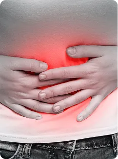
Here you will find informative articles about gut health, from possible causes and symptoms to strategies for relieving discomfort and sustainably supporting your digestive health.

Relevant Topics
Categories
Improve your gut health with our blog articles.
Everything about irritable bowel syndrome and other relevant topics.
Everything about other gastrointestinal diseases.
Low-FODMAP diet, gluten-free foods, and more!
Learn all about how the gut-brain axis works.
Here you can find out everything about the diagnostic procedures for gastrointestinal diseases.


Test your gut health with our self-test!

What are you waiting for?
We'll guide you step by step on your journey to noticeably fewer IBS symptoms.
The Cara-Newsletter

CE-certified medical device
data protection guaranteed
Cara Care. All rights reserved.